MightyMountTM Antifade Fluorescence Mounting Medium (aqueous)
Biological description
Overview
MightyMountTM Antifade Fluorescence Mounting Medium (aqueous) is an ideal formulation for prevention of photobleaching of fluorescent proteins and dyes during fluorescent imaging. It is easy to use with an ideal refractive index and provides effective prevention of photobleaching.
Figure 1. βIII-tubulin and Neurofilament light staining in rat cerebellum. Mounted using MightyMountTM Antifade Fluorescence Mounting Medium (aqueous).
4% PFA fixed 40µm horizontal rat brain sections were stained for βIII-tubulin (HB6639) and Neurofilament light (HB7266) with DAPI (HB0747) used as a nuclear counterstain. Sections were mounted using MightyMountTM Antifade Fluorescence Mounting Medium (aqueous). For a full protocol and more information on IHC(IF) please see our IHC(IF) protocol.
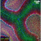
Figure 2. GFAP and NeuN staining in rat hippocampus. Mounted using MightyMountTM Antifade Fluorescence Mounting Medium (aqueous).
4% PFA fixed 40µm horizontal rat brain sections were stained for GFAP (HB8267) and NeuN (HB6498) with DAPI (HB0747) used as a nuclear counterstain. Sections were mounted using MightyMountTM Antifade Fluorescence Mounting Medium (aqueous). For a full protocol and more information on IHC(IF) please see our IHC(IF) protocol.
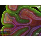
Figure 3. MBP and Neurofilament L staining in rat cerebellum. Mounted using MightyMountTM Antifade Fluorescence Mounting Medium (aqueous).
4% PFA fixed 40µm horizontal rat brain sections were stained for MBP (HB8014) and Neurofilament L (HB7266) with DAPI (HB0747) used as a nuclear counterstain. Sections were mounted using MightyMountTM Antifade Fluorescence Mounting Medium (aqueous). For a full protocol and more information on IHC(IF) please see our IHC(IF) protocol.
Figure 4. Antifade performance of MightyMountTM Antifade Fluorescence Mounting Medium (aqueous) compared to VECTASHIELD® and Fluoromount-G™.
Hello Bio MightyMountTM Antifade Fluorescence Mounting Medium (aqueous) shows strong antifade characteristics against a range of fluorophores and compares favorably against VECTASHIELD® and Fluoromount-G™. Method: HEK293T cells were cultured on coverslips then stained with DAPI (HB0747) and with a mouse anti-β-tubulin antibody (HB6491) with Alexa Fluor 488 or DyLight 550 conjugated secondary antibodies. Following mounting with either MightyMountTM Antifade Fluorescence Mounting Medium (aqueous), VECTASHIELD® or Fluoromount-G™, slides were imaged on a confocal microscope. Imaging was carried out as a timeseries at 100% laser power with the normalized intensity being calculated compared to the first exposure.
Figure 5. GluN1 and NeuN staining in rat cerebellum. Mounted using MightyMountTM Antifade Fluorescence Mounting Medium (aqueous).
Glyoxal fixed (9% glyoxal, 8% acetic acid, pH4) 40µm horizontal rat brain sections were stained for GluN1 (HB7535) and NeuN (HB6498) with DAPI (HB0747) used as a nuclear counterstain. Sections were mounted using MightyMountTM Antifade Fluorescence Mounting Medium (aqueous). For a full protocol and more information on IHC(IF) please see our IHC(IF) protocol.
Figure 6. βIII-tubulin and Neurofilament light staining in rat cortex. Mounted using MightyMountTM Antifade Fluorescence Mounting Medium (aqueous).
4% PFA fixed 40µm horizontal rat brain sections were stained for βIII-tubulin (HB6639) and Neurofilament light (HB7266) with DAPI (HB0747) used as a nuclear counterstain. Sections were mounted using MightyMountTM Antifade Fluorescence Mounting Medium (aqueous). For a full protocol and more information on IHC(IF) please see our IHC(IF) protocol.
Figure 7. βIII-tubulin and Neurofilament light staining in rat cerebellum. Mounted using MightyMountTM Antifade Fluorescence Mounting Medium (aqueous).
4% PFA fixed 40µm horizontal rat brain sections were stained for βIII-tubulin (HB6639) and Neurofilament light (HB7266) with DAPI (HB0747) used as a nuclear counterstain. Sections were mounted using MightyMountTM Antifade Fluorescence Mounting Medium (aqueous). For a full protocol and more information on IHC(IF) please see our IHC(IF) protocol.
Figure 8. βIII-tubulin and Neurofilament light staining in rat cerebellum. Mounted using MightyMountTM Antifade Fluorescence Mounting Medium (aqueous).
4% PFA fixed 40µm horizontal rat brain sections were stained for βIII-tubulin (HB6639) and Neurofilament light (HB7266) with DAPI (HB0747) used as a nuclear counterstain. Sections were mounted using MightyMountTM Antifade Fluorescence Mounting Medium (aqueous). For a full protocol and more information on IHC(IF) please see our IHC(IF) protocol.
Figure 9. MBP and Neurofilament L staining in rat dentate gyrus. Mounted using MightyMountTM Antifade Fluorescence Mounting Medium (aqueous).
4% PFA fixed 40µm horizontal rat brain sections were stained for MBP (HB8014) and Neurofilament L (HB7266) with DAPI (HB0747) used as a nuclear counterstain. Sections were mounted using MightyMountTM Antifade Fluorescence Mounting Medium (aqueous). For a full protocol and more information on IHC(IF) please see our IHC(IF) protocol.
Figure 10. Neurofilament L staining in rat dentate gyrus. Mounted using MightyMountTM Antifade Fluorescence Mounting Medium (aqueous).
4% PFA fixed 40µm horizontal rat brain sections were stained for Neurofilament L (HB7266) with DAPI (HB0747) used as a nuclear counterstain. Sections were mounted using MightyMountTM Antifade Fluorescence Mounting Medium (aqueous). For a full protocol and more information on IHC(IF) please see our IHC(IF) protocol.
Figure 11. MBP and Neurofilament L staining in rat cerebellum. Mounted using MightyMountTM Antifade Fluorescence Mounting Medium (aqueous).
4% PFA fixed 40µm horizontal rat brain sections were stained for MBP (HB8014) and Neurofilament L (HB7266) with DAPI (HB0747) used as a nuclear counterstain. Sections were mounted using MightyMountTM Antifade Fluorescence Mounting Medium (aqueous). For a full protocol and more information on IHC(IF) please see our IHC(IF) protocol.
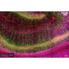
Figure 12. MBP and Neurofilament L staining in rat subiculum. Mounted using MightyMountTM Antifade Fluorescence Mounting Medium (aqueous).
4% PFA fixed 40µm horizontal rat brain sections were stained for MBP (HB8014) and Neurofilament L (HB7266) with DAPI (HB0747) used as a nuclear counterstain. Sections were mounted using MightyMountTM Antifade Fluorescence Mounting Medium (aqueous). For a full protocol and more information on IHC(IF) please see our IHC(IF) protocol.
Figure 1. βIII-tubulin and Neurofilament light staining in rat cerebellum. Mounted using MightyMountTM Antifade Fluorescence Mounting Medium (aqueous).
4% PFA fixed 40µm horizontal rat brain sections were stained for βIII-tubulin (HB6639) and Neurofilament light (HB7266) with DAPI (HB0747) used as a nuclear counterstain. Sections were mounted using MightyMountTM Antifade Fluorescence Mounting Medium (aqueous). For a full protocol and more information on IHC(IF) please see our IHC(IF) protocol.
Figure 2. GFAP and NeuN staining in rat hippocampus. Mounted using MightyMountTM Antifade Fluorescence Mounting Medium (aqueous).
4% PFA fixed 40µm horizontal rat brain sections were stained for GFAP (HB8267) and NeuN (HB6498) with DAPI (HB0747) used as a nuclear counterstain. Sections were mounted using MightyMountTM Antifade Fluorescence Mounting Medium (aqueous). For a full protocol and more information on IHC(IF) please see our IHC(IF) protocol.
Figure 3. MBP and Neurofilament L staining in rat cerebellum. Mounted using MightyMountTM Antifade Fluorescence Mounting Medium (aqueous).
4% PFA fixed 40µm horizontal rat brain sections were stained for MBP (HB8014) and Neurofilament L (HB7266) with DAPI (HB0747) used as a nuclear counterstain. Sections were mounted using MightyMountTM Antifade Fluorescence Mounting Medium (aqueous). For a full protocol and more information on IHC(IF) please see our IHC(IF) protocol.
Figure 4. Antifade performance of MightyMountTM Antifade Fluorescence Mounting Medium (aqueous) compared to VECTASHIELD® and Fluoromount-G™.
Hello Bio MightyMountTM Antifade Fluorescence Mounting Medium (aqueous) shows strong antifade characteristics against a range of fluorophores and compares favorably against VECTASHIELD® and Fluoromount-G™. Method: HEK293T cells were cultured on coverslips then stained with DAPI (HB0747) and with a mouse anti-β-tubulin antibody (HB6491) with Alexa Fluor 488 or DyLight 550 conjugated secondary antibodies. Following mounting with either MightyMountTM Antifade Fluorescence Mounting Medium (aqueous), VECTASHIELD® or Fluoromount-G™, slides were imaged on a confocal microscope. Imaging was carried out as a timeseries at 100% laser power with the normalized intensity being calculated compared to the first exposure.
Figure 5. GluN1 and NeuN staining in rat cerebellum. Mounted using MightyMountTM Antifade Fluorescence Mounting Medium (aqueous).
Glyoxal fixed (9% glyoxal, 8% acetic acid, pH4) 40µm horizontal rat brain sections were stained for GluN1 (HB7535) and NeuN (HB6498) with DAPI (HB0747) used as a nuclear counterstain. Sections were mounted using MightyMountTM Antifade Fluorescence Mounting Medium (aqueous). For a full protocol and more information on IHC(IF) please see our IHC(IF) protocol.
Figure 6. βIII-tubulin and Neurofilament light staining in rat cortex. Mounted using MightyMountTM Antifade Fluorescence Mounting Medium (aqueous).
4% PFA fixed 40µm horizontal rat brain sections were stained for βIII-tubulin (HB6639) and Neurofilament light (HB7266) with DAPI (HB0747) used as a nuclear counterstain. Sections were mounted using MightyMountTM Antifade Fluorescence Mounting Medium (aqueous). For a full protocol and more information on IHC(IF) please see our IHC(IF) protocol.
Figure 7. βIII-tubulin and Neurofilament light staining in rat cerebellum. Mounted using MightyMountTM Antifade Fluorescence Mounting Medium (aqueous).
4% PFA fixed 40µm horizontal rat brain sections were stained for βIII-tubulin (HB6639) and Neurofilament light (HB7266) with DAPI (HB0747) used as a nuclear counterstain. Sections were mounted using MightyMountTM Antifade Fluorescence Mounting Medium (aqueous). For a full protocol and more information on IHC(IF) please see our IHC(IF) protocol.
Figure 8. βIII-tubulin and Neurofilament light staining in rat cerebellum. Mounted using MightyMountTM Antifade Fluorescence Mounting Medium (aqueous).
4% PFA fixed 40µm horizontal rat brain sections were stained for βIII-tubulin (HB6639) and Neurofilament light (HB7266) with DAPI (HB0747) used as a nuclear counterstain. Sections were mounted using MightyMountTM Antifade Fluorescence Mounting Medium (aqueous). For a full protocol and more information on IHC(IF) please see our IHC(IF) protocol.
Figure 9. MBP and Neurofilament L staining in rat dentate gyrus. Mounted using MightyMountTM Antifade Fluorescence Mounting Medium (aqueous).
4% PFA fixed 40µm horizontal rat brain sections were stained for MBP (HB8014) and Neurofilament L (HB7266) with DAPI (HB0747) used as a nuclear counterstain. Sections were mounted using MightyMountTM Antifade Fluorescence Mounting Medium (aqueous). For a full protocol and more information on IHC(IF) please see our IHC(IF) protocol.
Figure 10. Neurofilament L staining in rat dentate gyrus. Mounted using MightyMountTM Antifade Fluorescence Mounting Medium (aqueous).
4% PFA fixed 40µm horizontal rat brain sections were stained for Neurofilament L (HB7266) with DAPI (HB0747) used as a nuclear counterstain. Sections were mounted using MightyMountTM Antifade Fluorescence Mounting Medium (aqueous). For a full protocol and more information on IHC(IF) please see our IHC(IF) protocol.
Figure 11. MBP and Neurofilament L staining in rat cerebellum. Mounted using MightyMountTM Antifade Fluorescence Mounting Medium (aqueous).
4% PFA fixed 40µm horizontal rat brain sections were stained for MBP (HB8014) and Neurofilament L (HB7266) with DAPI (HB0747) used as a nuclear counterstain. Sections were mounted using MightyMountTM Antifade Fluorescence Mounting Medium (aqueous). For a full protocol and more information on IHC(IF) please see our IHC(IF) protocol.
Figure 12. MBP and Neurofilament L staining in rat subiculum. Mounted using MightyMountTM Antifade Fluorescence Mounting Medium (aqueous).
4% PFA fixed 40µm horizontal rat brain sections were stained for MBP (HB8014) and Neurofilament L (HB7266) with DAPI (HB0747) used as a nuclear counterstain. Sections were mounted using MightyMountTM Antifade Fluorescence Mounting Medium (aqueous). For a full protocol and more information on IHC(IF) please see our IHC(IF) protocol.
Biological Data
Application notes
Protocol for use of mounting media
IHC(IF)
Mount sections onto subbed or charged microscope slides and air dry (in the dark) until sections are moist but all excess liquid has evaporated
Add a few drops of mounting media around the sections (around 50µl but this will depend on the number and thickness of sections) and slowly lower the coverslip from one end of the slide to the other being careful to avoid creating any bubbles.
Use clear nail varnish to seal the edges of the slide to avoid movement during imaging and stop evaporation.
For more information on IHC(IF) including tips on how to mount sections, please see our IHC(IF) protocol
ICC
Add a drop of mounting medium (Around 5µl for a 10mm and 15µl for a 22mm coverslip) to a standard microscope slide.
Briefly rinse the coverslip in dH2O before placing face down into the drop of mounting medium being careful not to introduce bubbles.
Use clear nail varnish to seal the edges of the coverslip to avoid movement during imaging and stop evaporation.
For more information on ICC please see our ICC protocol
Solubility & Handling
Storage instructions
+4°C or -20°C long-term. Protect from light.
Important
This product is for RESEARCH USE ONLY and is not intended for therapeutic or diagnostic use. Not for human or veterinary use.


.")
.")
.")
 compared to VECTASHIELD® and Fluoromount-G™.")
.")
.")
.")
.")
.")
.")
.")
.")